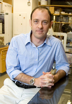
Andres Leschziner
What type of research might appeal to a young scientist whose interests span the elegance of chemistry and the excitement of biology? For Andres Leschziner, structural biology was the compelling choice. A new MCB-assistant professor, Leschziner views structural biology-which reveals the architecture and shape of macromolecules-as fertile ground for discovery.
Pushing Boundaries in 3D-Electron Microscopy
Leschziner brings with him top-flight expertise in three-dimensional electron microscopy (3D-EM), a tool that images macromolecules using streams of electrons instead of light. The technology is particularly well suited for big, flexible molecules that adopt multiple conformations, Leschziner says. As such, it can expose hidden territory in molecular biology. Scientists now recognize that for many macromolecules, flexibility is the rule, not the exception. Moreover, that flexibility has biological significance-molecules behave differently when they adopt one conformation over another. But constrained by available methods, scientists have been challenged in their efforts to study flexibility at high resolution, particularly in large molecules. 3D-EM will change that, Leschziner predicts. But he concedes the technology’s full potential has yet to be realized. “Right now it’s very specialized,” he says. “We want to advance 3D-EM to the point that it becomes routine in biological research.”
Heading North from Argentina
Until his college years, Leschziner lived in Argentina; where his grandparents resettled after fleeing Europe during WWII. His father’s parents endured what he calls a “movie-like get-away,” from Germany in 1938; escaping first to the Netherlands, then to Rio de Janeiro, where they were briefly jailed before continuing to Santiago, Chile, and then east to Argentina. His mother was born in a Swiss refugee camp during the war, and afterwards lived in Paris, until she and her family left for South America in 1953.
Leschziner completed two years of college in Argentina, and then emigrated to Canada, picking up English and an undergraduate degree in biology, from McGill University in 1993. From there, he left for Yale, where his interest in structural biology was sparked. Working with his advisors Thomas Steitz and Nigel D. F. Grindley, Leschziner developed a method that allowed him to determine how a pair of protein dimers that cut and splice DNA come together during DNA recombination. Scientists had already discerned the complex’s shape using X-ray crystallography but they hadn’t been able to figure out how its components were oriented during recombination. To solve that problem, Leschziner took a unique approach: he created a system that allowed him to engineer all the plausible orientations, and then he back-tracked through them to expose the correct one. “What was creative about this work is that it used biochemistry to tackle a “structural” question that crystallography hadn’t been able to answer at the time,” Leschziner asserts. “So, my experiments were both heavily informed by and designed to yield structural information. But I wasn’t a structural biologist yet.”
That changed when Leschziner headed for Berkeley to pursue post-doctoral training in structural biology with Professor Eva Nogales in a laboratory that-much to his liking-was just two years old. Leschziner says he was attracted to the notion of working in a small, growing laboratory because it gave him the opportunity to interact closely with his colleagues. At Berkeley, he turned his attention to 3D-EM, viewing it as the most exciting thing happening in structural biology at the time. A relatively new technology, 3D-EM collects images that contain detailed information about a molecule’s internal structure. By figuring out how these different views relate to each other in space, scientists can reconstruct the molecule in three dimensions.
3D-EM fills a gap in structural biology, because it works with large molecules under physiological conditions; something that other techniques in the field-namely X-ray crystallography and nuclear magnetic resonance imaging-can’t achieve. To its detriment, however, it requires intensive computations, and apart from a few “spectacular” exceptions, Leschziner says, it can’t yet achieve atomic resolution.
Filling the Missing Cone
In short, 3D-EM was then-as it is now-a field imbued with potential. At Berkeley, Leschziner used it to study chromatin remodeling complexes, which are macromolecules that help cells deal with the tight packaging of DNA in their nuclei. But he ran into a barrier: Scientists trying to generate initial 3D reconstructions with the technology often encounter a blind spot called the “missing cone.”
Leschziner set out to solve the problem with a new approach that he called orthogonal tilt reconstruction (OTR). His method fills the missing cone by collecting data at -45 and +45 degrees (two orthogonal tilts); a geometry that allows users to create reconstructions without the missing information, thus eliminating the need for decisions that could bias results. Leschziner went on to use OTR to study conformational flexibility in a chromatin remodeler called RSC, from the yeast S. cerevisiae. Meanwhile, the OTR method-viewed as a key advance for the field-launched his growing reputation within the 3D-EM community.
Leschziner recently hired a computer programmer to write software that interfaces between users and underlying scripts. Ideally, scientists will be able to run OTR by pushing a few buttons, without worrying about the requisite computations, he says. “I’d be very pleased if the method became established as a way to generate initial reconstructions in a fully automated way.”
Meanwhile, visualizing flexibility at high resolution remains a key goal. It’s difficult to understate how hard it is to solve large macromolecule structures, however, particularly if they lack symmetry. Viruses, for instance, are highly symmetrical particles that in some cases can be reconstructed to high resolution with 3D-EM in a matter of days. More complex, asymmetrical structures take far longer.
Though he’s best known for his work on 3D-EM, Leschziner remains a biologist at heart. “If things go well I hope to have made a major contribution towards our understanding of how chromatin remodeling works,” he says. Along those lines, he plans to continue his ongoing collaborations with Bradley Cairns, a professor in oncology at the Huntsman Cancer Institute at the University of Utah, and also with Carl Wu, a laboratory chief in biochemistry and molecular biology at the National Cancer Institute. Closer to home, he’s also engaged in collaborations with MCB professor Tom Maniatis, investigating chromatin remodeling in larger biological contexts, he says.
Leschziner arrived at Harvard this year together with his wife, Samara (Sam) Reck-Peterson, a junior faculty member in cell biology at Harvard medical school. “There’s a sense of excitement for the science going on at Harvard and in Boston in general that’s hard to resist” he says. “The quality of my colleagues and that of students and post-docs was also a big attraction. As for MCB, I find the breadth of the department very appealing. I find that my sense of amazement for biology-the one that got me into science to begin with-has been rekindled by the variety of things I’m exposed to here on a regular basis. In deciding to come here I was attracted to the idea that having such diverse colleagues can take my research into unexpected directions.”