After nearly 20 years of meticulous research, a team of Harvard scientists has published findings in Nature Neuroscience (PDF) that fundamentally challenge how we think about brain development. The study reveals that neural activity—the function of the brain—actually dictates how neurons wire themselves together, rather than the reverse.
“The usual way of thinking about this is that the structure of the brain explains its function,” said Jeff Lichtman, the Jeremy R. Knowles Professor of Molecular and Cellular Biology. “This is work that shows that the function of the brain generates structure.”
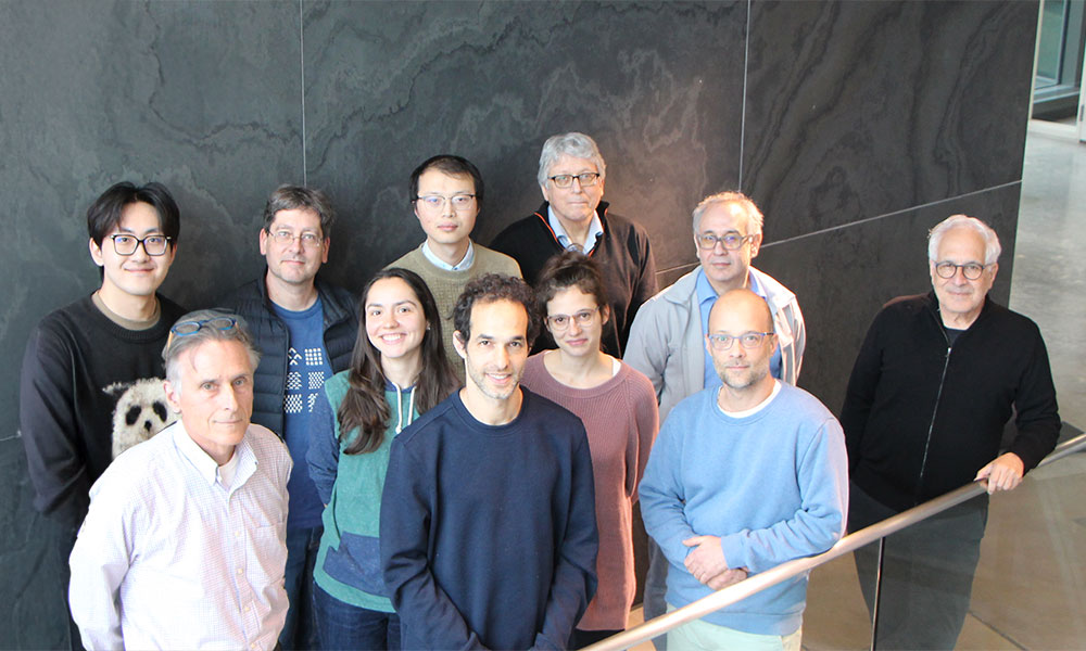
The discovery emerged from an ambitious collaboration that brought together multiple cutting-edge technologies and spans from some of Lichtman’s earliest work at Harvard to the present day. Ryan Draft, who was among Lichtman’s first PhD students after he joined Harvard in 2004, carried out the initial studies that would eventually become foundational for this landmark project a decade later. Draft’s work marked the start of a long investigation into how the neuromuscular circuit assembles itself from birth through adulthood. Draft’s earliest studies would only later be connected to what the team discovered from newborn electron microscopy (EM) data and the developmental connectomic work eventually led by Yaron Meirovitch.
A Two-Decade Scientific Journey
The project’s remarkable timeline reflects both the technical challenges involved and the care taken to ensure the findings were robust and comprehensive. Draft began the work during his PhD between 2007 and 2010, using newly developed Brainbow technology—a method invented by Lichtman and colleagues that labels individual neurons with distinct fluorescent colors, allowing researchers to track them through complex neural networks.
“We were really interested in using the neuromuscular junction as a model system, because it’s simple compared to any other part of the brain, and super accessible,” said Draft, now an academic advisor for Harvard’s undergraduate neuroscience program. The neuromuscular junction is where motor neurons connect to muscle fibers, providing an ideal window into fundamental principles of neural development.
Draft’s early fluorescent microscopy work revealed surprising patterns in how neurons competed for synaptic connections during development, but the resolution wasn’t sufficient to see what was happening at birth, when the circuits are most densely interconnected. That’s where the EM work began. Around 2013, Kai Kang, former postdoc in the Lichtman lab and co-first author on this paper, started the herculean task of preparing an entire newborn muscle for synaptic-resolution reconstruction of all motor neurons and their connections to muscle fibers using serial section electron microscopy.
When Meirovitch joined Harvard in 2018, he first undertook the challenge of reconstructing the newborn connectome from this muscle. Trained as a PhD student at the Weizmann Institute of Science in Israel studying organizational principles of the motor system with Tamar Flash, Meirovitch arrived with a deep interest in how structured patterns emerge from neural activity – an intellectual thread that aligned naturally with the project’s central question. This work required both machine learning and painstaking manual reconstruction by Meirovitch and a team of undergraduate interns, who traced extremely fine axonal branches through hundreds of neuromuscular junctions (NMJs) and thousands of serial EM sections. This newborn dataset proved to be one of the most difficult reconstruction tasks attempted in connectomics at that time.
As the project expanded to include multiple developmental muscles, Juan Carlos Tapia – a long-time collaborator who had worked with Lichtman before his move to Harvard – returned to contribute essential biological expertise, including helping establish the existence of multiple NMJs on single fibers. Together with additional computational help from Ju Lu, Lichtman’s first PhD student at Harvard, the team found that not only can muscle fibers have more than one NMJ in the adult, but that this arrangement is far more abundant at birth—and that, through maturation, entire NMJs with all their axonal inputs are selectively eliminated.
“At some point, I went to Jeff’s office and showed him a plot,” Meirovitch recalled. “He looked at the data and saw exactly what Ryan had observed with Brainbow years earlier. That was the turning point – the moment we began to bridge decades of research to see the full picture.”
Function Before Structure: A Paradigm Shift
The team’s central finding challenges a fundamental assumption in neuroscience. Using the neuromuscular system as their model, they discovered that even before mice can move their muscles at birth, the neural activity associated with planned movements is already present upstream in the motor system—and this activity shapes how the nervous system ultimately wires itself to control those muscles.
“What we see is that neural activity propagating from one region in the nervous system to another is not waiting for the structure to form at its target,” Meirovitch explained. “The activity is already there, and it is shaping the structure as it emerges. The function truly comes before the structure.”
The study tracked neural development from birth through adulthood, documenting how multiple motor neurons initially compete to innervate each muscle fiber, but eventually only one connection survives. Crucially, the researchers found that neurons firing in synchrony—at the same time—were more likely to maintain their connections and continue competing, while neurons with mistimed activity were eliminated more quickly.
“People have said for decades that neurons that fire together wire together,” Meirovitch said, referring to a principle proposed some 60 years ago. “But now we see a concrete biological mechanism for that idea, both at the single-synapse and the circuit levels, and its implementation in the natural setting of coordinate movement.”
Unexpected Discovery: Long-Distance Competition
The team also made a surprising discovery that even Lichtman initially doubted: some muscle fibers have multiple neuromuscular junctions separated by up to a millimeter—and these distant sites can compete with each other for survival using propagating action potentials on the muscle fibers. This long-distance competition was not previously known to occur consistently in adult mice.
“If their action potentials collide, the junctions never really ‘see’ each other,” Meirovitch explained. “But if the signals are sufficiently out of sync, they pass through, and the junctions become ‘aware’ of their rivals. That initiates a competition – one that typically ends with the weaker, mistimed NMJ being eliminated entirely.”
“Unexpectedly, we found that at birth, many more muscle fibers host multiple NMJs. During maturation, these junctions gradually disappear based on their timing relationships and the toll of synaptic activity on the motor neurons,” he said. This mechanism offers an elegant solution to how neural circuits become more efficient during development—by removing redundant connections while preserving those that work together in coordinated patterns.
Tapia’s biological work was key in this part of the project, helping establish the existence and developmental fate of these multiple NMJ sites. His contributions underscored the historical depth of the project, having collaborated with Lichtman decades earlier and returning to help resolve one of its most surprising findings.
A Testament to Scientific Persistence
For Lichtman, this paper represents the culmination of a research trajectory that began during his own graduate studies decades ago. “This is the subject I began working on as a graduate student, and this paper finally puts it all together,” he said. “For me, personally, it’s an extremely important piece of work.”
Rather than publish the findings piecemeal, the team waited until they could present a comprehensive picture. “We had three or four different sets of studies, each of which was quite mysterious,” Lichtman explained. “We were delaying publishing because we weren’t actually sure how they fit together. It was only in the last go around that it was possible to see this was all looking at the same gigantic elephant, just from different angles.”
The study showcases the power of combining complementary technologies—Brainbow’s ability to identify individual neurons over large areas, and electron microscopy’s capacity to resolve fine structural details—to answer fundamental biological questions. It also demonstrates how some scientific questions require extraordinary patience and collaboration to answer properly.
“It’s kind of a tour de force,” said Draft, reflecting on the project’s scope. “It’s really a big deal to show that there’s a lot to be learned from looking at the development of these networks over time, and now we have some techniques to do it.”
The findings may have implications beyond the neuromuscular system, potentially revealing principles that operate throughout the nervous system at different developmental stages and timescales. Understanding how neural circuits self-organize to create the precise connections needed for behavior represents a fundamental question in neuroscience—one this team has spent two decades working to answer.
![]() (PDF)
(PDF)