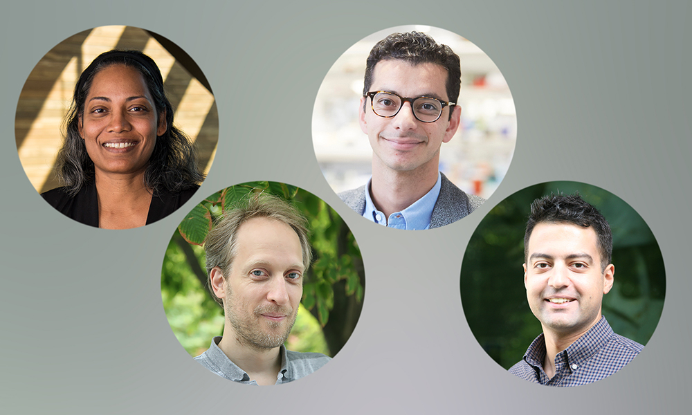

Several MCB-affiliated faculty members have been selected as recipients of the Star-Friedman Challenge for Promising Scientific Research, one of Harvard’s most prestigious internal funding competitions. The awards, administered by the Faculty of Arts and Sciences Office of Research Administration, support bold, innovative projects that push investigators into new scientific directions.
The 2026 MCB-affiliated recipients and their projects are:

Doeke Hekstra, Associate Professor of Molecular & Cellular Biology and of Applied Physics (SEAS), and Victoria D’Souza, MCB Professor, “Uncovering the Physiological Shapes of Proteins by Orienting Them Electrically”
Many of biology’s most important molecules—viral replication machinery, proteins transcribing DNA into mRNA, antibodies, and other large protein- or protein–nucleic acid complexes (here “proteins” for short)—do not adopt a single rigid shape. Instead, they shift among shapes (conformations) in the watery solutions in which they work. These shapes determine biological function and drug binding. Currently, methods for determining these shapes at atomic resolution require either crystallizing the protein for X-ray crystallography or freezing it for cryogenic electron microscopy (cryo-EM). Unfortunately, both during crystal formation and during cooling the equilibrium between shapes often shifts, such that the observed shapes are not necessarily the ones important inside the human body.
An important example is the Human Immunodeficiency Virus-1 (HIV-1) initiation complex (IC) – a 2:2 protein-RNA complex forming a critical step in viral reproduction. In work by D’Souza, cryo-EM reveals conformations of the IC, of which only one is consistent with small-angle X-ray (SAXS) solution scattering data. Indeed, SAXS provides unique access to the solution-state behavior of large proteins but contains only limited information because the signal is an average over all orientations found in a sample. That averaging is often the difference between “compatible with many models” and “the data clearly prefer one model.”
Listening to a talk on RNA dynamics last year, Hekstra wondered how, in 1952 (!), Franklin and Gosling were able to collect the famous “Photograph 51”, leading to the elucidation of the first structure of DNA, including the 3.3 Å spacing of its base pairs, while such structural detail is barely achievable with solution X-ray scattering data nearly 75 years later. The key difference is that the DNA molecules were all aligned, unlike the usual random (isotropic) orientations found in solution.
In their project, the team endeavors to orient protein molecules in solution for just long enough to take such snapshots by developing electric-field oriented wide-angle X-ray scattering (eWAXS): a method that aligns molecules in solution using brief electric-field pulses, allowing researchers to recover otherwise averaged structural information. If successful, eWAXS will become a platform for determining which conformations dominate under physiological conditions for large biomolecular complexes—precisely the most important, yet inaccessible regime. The method could reshape how researchers validate drug targets, antibody binding modes, and the interplay between the binding partners of large protein and ribonucleoprotein assemblies.
“Victoria and I are very grateful for the support from the Star-Friedman Challenge,” Hekstra said. “This project really brings together engineering, physics, biochemistry, and virology in a novel way—both for us and for the field. We hope to open the door to critical new information on proteins that will help us better understand how they work and how to intervene to treat disease.”
Maxim Prigozhin, Assistant Professor of Molecular & Cellular Biology and of Applied Physics, “Revealing Protein Mechanics Using High-Pressure Cryo-Electron Microscopy”
Proteins are not static structures — they continuously shift through multiple conformational states as they carry out their functions inside cells. Yet most structural biology methods capture only the most energetically stable shape, leaving rarer but functionally critical conformations invisible. Prigozhin’s project asks whether hydrostatic pressure can coax proteins into these hidden states, making them detectable by modern cryo-electron microscopy (cryo-EM).
The implications are broad. At a fundamental level, mapping the full conformational landscape of a protein will reveal how mechanical forces propagate through protein structures and illuminate allosteric networks — the internal communication pathways that regulate protein activity. In the context of cellular imaging, structures obtained under pressure could also improve template matching in cryo-electron tomography, where proteins embedded in the crowded cellular environment may adopt shapes that differ from those seen in purified samples.
The work also has potential significance for drug discovery. Many small-molecule drugs bind to transient “cryptic pockets” that are only exposed in rare protein conformations. By systematically revealing these states, the project may open new avenues for pharmacological targeting. More broadly, the research aims to establish high-pressure cryo-EM as a generalizable tool for probing the hidden structural repertoire of proteins.
“My lab and I are very grateful to the donors, Mr. Star and Mr. Friedman, for establishing this award and for funding our work,” Prigozhin said. “We really appreciate the opportunity to jump-start this high-risk, high-reward project that we’d struggle to get off the ground without this generous seed support. We also want to congratulate all the fellow awardees— we are humbled to be included in this 2026 cohort of winners.”
Ahmad “Mo” Khalil, MCB Professor, and Hok Lam and Kathleen Kam Wong Professor of Bioengineering, and Manoj Duraisingh, John LaPorte Given Professor of Immunology and Infectious Diseases
“Engineering Red Blood Cells for Disease Detection: PATROL-RBCs (Programmable Antigen-Triggered Recognition and Organism-wide Logger–RBCs)”
Many diseases — including cancer and chronic inflammatory conditions — are defined by rare or spatially distributed pathological cells that are difficult to detect using conventional diagnostics. Current approaches rely on transient biomarkers or single-time-point measurements, and therefore miss the dynamic cell-cell interactions that occur within tissues. Duraisingh and Khalil’s project proposes a fundamentally new diagnostic platform: engineering red blood cells (RBCs) to patrol the body, recognize disease-associated cells through direct contact, and record those encounters in a stable, measurable way.
Red blood cells are uniquely suited to this challenge. As the most abundant cells in the body, they traverse every vascular bed continuously over their roughly 120-day lifespan, sampling physiological and pathological environments throughout the organism. Unlike synthetic nanoparticles, RBCs are living cells with defined membrane architecture capable of hosting sophisticated molecular functions — yet they are enucleated and immunologically quiet, minimizing the risk of uncontrolled gene expression or immune rejection.
The project brings together two complementary research programs. The Duraisingh laboratory has deep expertise in RBC biology through years of studying malaria, including the development of immortalized erythroid lines and in vitro culture systems that produce engineered RBCs closely resembling healthy donor cells. The Khalil laboratory is a leader in synthetic biology and has pioneered programmable receptor systems — including SNIPRs (synthetic intramembrane proteolysis receptors) — that convert cell-cell contact into defined intracellular outputs. Together, the two groups aim to endow RBCs with synthetic sensing and recording capabilities, creating what they term PATROL-RBCs: a long-lived, circulating diagnostic platform capable of logging encounters with pathological cells directly within the body’s own physiological environment.
“I am deeply grateful to the Star-Friedman Challenge Program for supporting and enabling investigators to take on new research directions, and for recognizing the exciting potential of applying synthetic biology (our expertise) to red blood cells (the expertise of the Duraisingh lab) to develop a new paradigm of blood-based diagnostics,” said Khalil.
Established in 2013 through a gift from James A. Star and expanded in 2018 through a gift from Joshua and Beth Friedman, the Star-Friedman Challenge funds projects across the life, physical, and social sciences. The Star-Friedman Challenge has supported more than 75 projects at Harvard since its founding.