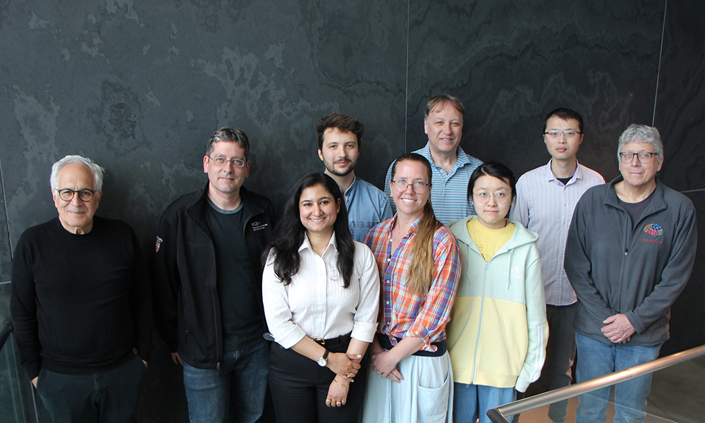
Since 2018, the Lichtman Lab has been painstakingly mapping every cell and synapse in a tiny brain sample from a human patient. Although the sample represents only one millionth of the volume of a human brain, mapping it in such detail generated 1.4 million gigabytes of data. To manage such a large dataset, the Lichtman Lab teamed with Google’s connectomics research group. The collaboration yielded the largest connectomics map of a human sample produced so far. This week, the project is being capped off with a paper in Science. Data from the project has been made available online for other researchers to peruse or analyze.
Seeking insight into the human brain’s microstructure was a strong motivator for the team behind the new paper. “All of the thousands of brain cells and tens of millions of synaptic connections identified in our study would show up on a standard MRI scan as a single pixel!” says co-first author Alexander Shapson-Coe, who was a postdoc in the Lichtman Lab at the time of the study.
In their map, the co-authors saw several unexplained phenomena. Many pairs of neurons in the sample had extremely strong connections, with dozens of synapses connecting the same two cells. Such strong connections have not been reported in mouse brains. They also identified pairs of triangular cells pointing in opposite directions and “whorls” or “spools” of axons, where the axons would form complex curlicue shapes. The researchers don’t know how any of these structures influence function. This paper is only a first glimpse.
“We provide a lot of basic measurements for the distribution of different cell types and synapses in the sample”, says research scientist Daniel Berger of the Lichtman Lab, who is one of the co-first authors on the paper. “It establishes some ground rules for the human brain, with the caveat that it’s from an epilepsy patient, so we will need some other samples.” Connectomic studies of even larger samples of human brain tissue are already in the works.
Berger adds that this particular study came about because of a series of lucky coincidences. The first was that collecting an excised bit of tissue from a surgery on an epileptic patient yielded a sample, nicknamed “H01”, which was of high enough quality to undergo intense connectomic analysis. The second was Shapson-Coe joining the Lichtman Lab and eagerly pushing for imaging and studying the sample. The third was that Google’s connectomics group was interested in a new data set and able to help store and process the data.
“We (the Connectomics Group at Google Research) started collaborating with Professor Lichtman all the way back in 2018,” says Google staff research scientist and co-first author Michal Januszewski. “My expertise was in analyzing the largest connectomics datasets available at the time—about 2% the size of the H01 sample. Our group was looking to scale up our methods, and the Lichtman lab had the technology and know-how to acquire the necessary data.”
The co-authors are proud of their accomplishment and look forward to future findings. “To image a piece of human brain tissue, with a size on the order of millimeters, and a resolution on the order of nanometers, is unprecedented, and we have only scratched the surface in terms of biological analyses of the dataset,” says Shapson-Coe. “We have developed tools to enable the exploration and analysis of this dataset by any interested individual or group; I expect the most notable discoveries from this dataset are yet to be made!”


