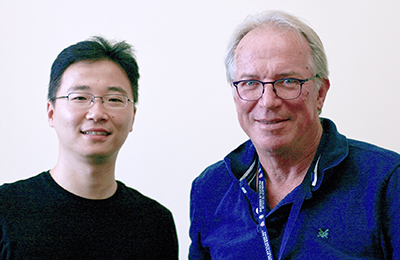
Tao Fang (l) and Hidde Ploegh
Brain is arguably the most complicated biological tissue. Many different cell types are intertwined and interconnected. Moreover the number of different cell types in the brain is vast and poorly understood. Molecular markers exist for many of these cell types and more are being uncovered at a rapid pace, thanks to the development of single-cell sequencing approaches. However, it is difficult to graft these molecular identities onto the cells revealed in connectomic studies, which rely heavily on the electron microscopy of an intact brain.
To superimpose (i.e. paint) the black-and-white EM images obtained in serial section electron microscopy with colors that signify their molecular identity, this work describes a new approach to correlate fluorescence light microscopy with electron microscopy (i.e. CLEM). Fluorescently tagged antibodies do provide easy-to-get labels for localizing many different molecular moieties in the same sample. But immunostaining requires detergents to create holes in cell membranes that invariably compromise the quality of electron microscopy-based ultrastructure which prevent connectomic analysis. In search of immunofluorescence staining agents that might be permeable without detergents we examined nanobodies. Nanobodies, which are lab-made fragments of heavy-chain-only antibodies produced in sharks, camels and related animals are essentially downsized antibodies. They only have 1/10 molecular weight of full-size antibodies; but, like a typical antibody, nanobodies are able to bind selectively to specific antigens. We tapped the size dividend of nanobodies and developed NATIVE (Nanobody-Assisted Tissue Immunostaining for Volumetric Electron microscopy), a tissue processing and staining method for large-scale CLEM imaging without compromising the ultrastructure.
To perform NATIVE, the nanobodies to brain antigens were conjugated with organic fluorophores and added to a paraformaldehyde-fixed tissue. We found that without any permeabilization agent, the cytoplasm of the cells and processes were readily accessible to the fluorescent nanobodies. Because of this, we could do fluorescence confocal imaging on a tissue sample and then prepare the same sample for electron microscopy staining and imaging. High ultrastructural quality is preserved in NATIVE allowing us to trace out neural circuits with the color information of cellular identities superimposed from the confocal images onto the electron microscopy. Moreover, their small size allows them to penetrate deeper into volumetric samples than regular antibodies.
Since NATIVE fully decouples immunofluorescence from the subsequent EM staining, light and electron microscopy can each operate without compromise. It is likely therefore that other fluorescence imaging techniques such as light sheet or super-resolution techniques can also be coupled to NATIVE. We expect that NATIVE will also allow the exploration of other biological samples bedsides brain, with an unprecedented multiplicity of markers, resolution and scale.
by Xiaotang Lu and Jeff Lichtman


