Small transparent animals like zebrafish and C. elegans have long been heralded for their accessibility to optical methods to manipulate and monitor neuronal function. A major shift in these approaches occurred when fast microscopes and efficient data analysis began to allow whole-brain imaging.
Several years ago, researchers in Aravi Samuel’s lab and their collaborators started building the tools needed for whole-brain imaging in C. elegans. Mei Zhen, Vivek Venkatachalam, and members of the Samuel lab assembled the tools and microscopes that were needed to begin whole-brain calcium imaging in behaving worms. Vlad Susoy, a postdoc in the Samuel lab, has now applied these methods to examine brain-wide mechanisms at single-neuron resolution across an entire behavioral repertoire that a brain organizes, the mating behavior of male worms. This work has recently been published in Cell.
“Our basic technology is a tracking microscope. I got into science as an undergraduate working with Howard Berg, who built the first tracking microscope to follow the activity of a single-celled ‘brain’ during an unrestricted behavior. That was how Howard unraveled the algorithmic structure of E. coli chemotaxis from stimulus input to motor output. I’ve always wanted to do something like that in my own lab, but with an animal, and now we have. Vlad measured every sensory neuron, interneuron, and motor neuron in the brain of the mating male throughout a behavioral trajectory and discovered many of the rules of the game,” says Aravi Samuel, Professor of Physics.
Recording from an entire brain was only a part of the challenge, another part was creating an experimental regime where the animal could interact with a diverse set of naturalistic stimuli as opposed to delivering artificial stimuli to fixed animals. To tackle this challenge the authors turned to nature itself and focused on a behavior that the animal wants to perform on its own – mating. Simply placing two nematodes together triggers mating, arguably their most complex natural behavior.
“Like many natural behaviors in mammals and invertebrates, nematode mating is goal-directed and unfolds in a variable sequence of many stereotyped behavioral steps. Each of these steps is driven by complex sensory stimuli that act through multiple sensory modalities induced by highly dynamic interactions between two mating partners,” says Vlad Susoy.
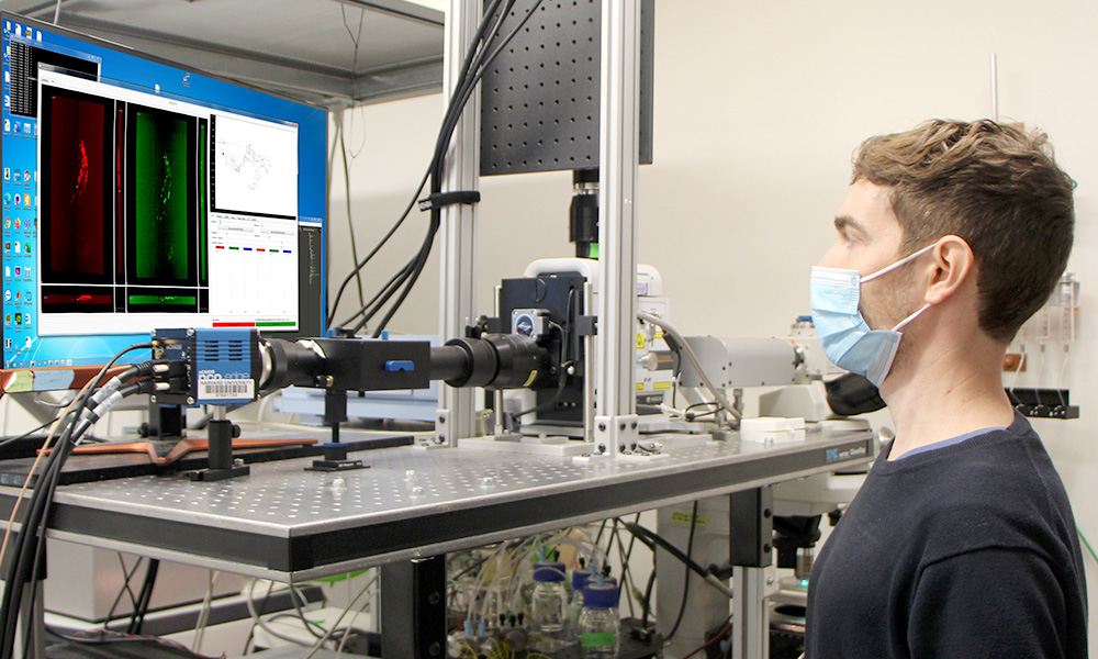
With a custom tracking microscope the researchers simultaneously recorded the behavior and neuronal activity in freely behaving nematodes from beginning to end of mating. When they compared brain-wide activities across different steps of mating, the researchers saw that the brain operates in the same way between instances of the same behavioral step whether recurring in one animal or occurring in different animals. This similarity was so pronounced, that it was possible to predict the behavior of one animal from the neuronal activity of other animals. In contrast, the brain operates differently between different steps of mating. When the brain-wide activity was compared across different steps of mating, unique neuron-neuron correlations specific to each behavioral step emerged. What causes these diverse context-specific correlations between neurons? The authors argue that it is the performance of behavior itself. Each of the many steps during mating presents different stimulus patterns. These stimulus patterns act on subsets of neurons, starting with the sensory neurons and create context-dependent interactions in the brain. The diversity of these interactions underlies the diversity of behavioral responses in an entire behavior. Remarkably when looking at the activity of each neuron relative to all steps of mating behavior, virtually every neuron in the circuit emerged as functionally unique. By closely looking at the neurons’ activities and mapping them onto the diagram of physical connections between neurons the researchers in Samuel’s lab could identify several mechanisms that the brain uses to process the information from the environment in a context-specific way to make decisions. The authors conclude that brain-wide dynamics during a natural behavior are not a phenomenon of an isolated nervous system but are shaped by the flow of sensory inputs and motor actions as behavior unfolds. Behavioral dynamics must be integrated with neuronal dynamics and wiring to understand the full range of brain function.
In the MCB Department at Harvard, Florian Engert’s lab has long applied whole-brain imaging to explore a range of different questions in the behaviors of zebrafish larva.
“In this study, Vlad and Aravi have recorded from most neurons in the worm’s brain throughout a complex behavioral sequence. In doing so they have not only shed light on how motor sequences can be generated by distributed networks of neurons, they also have passed a milestone in neuroscience that is still a very distant goal for the rest of the neuroscience community”, says Florian.
“While larval zebrafish, for example, afford in principle the possibility to record from all neurons in a behaving animal, the harsh reality is that this goal can only be met with quite serious caveats: in all preparations there is always a significant part of the cells that cannot be imaged, either because of labeling issues or compromised physical access, and the temporal resolution is always far away from the desired precision to faithfully report individual spikes. As thus, this project truly presents a milestone that allows us now to check how far such complete data sets can take us in updating our understanding of brain function. I believe there are many more very interesting stories awaiting just around the corner”.
By Vlad Susoy and Aravi Samuel


